
Hello, I'm Annemarie
I am a NY-based illustrator, animator, and interactive game designer. With expertise in medical illustration and biological anthropology, I bridge the gap between science and accessible knowledge through visually striking and scientifically accurate illustrations and animations.

EDUCATION
ROCHESTER INSTITUTE OF TECHNOLOGY
MFA Medical Illustration
2022 – 2025 (Expected)
Thesis: Creating a 3D Interactive Module for Teaching the Stages of Human Decomposition
Â
UNIVERSITY OF TENNESSEE KNOXVILLE
MA Biological Anthropology
2014 – 2017
Thesis: Geographic Information Systems and Predictive Modeling of Body Disposal Sites
Â
FORDHAM UNIVERSITY
BA Art History
BA Anthropology
2009 – 2013
EXPERIENCE
DEVELOPMENT & ADMIN MANAGER
Greene Land Trust
Oct 2024 – Present
Â
FREELANCE ILLUSTRATOR
Rochester Institute of Technology
PHT180
Apr 2023 – May 2024
Â
GRADUATE ASSISTANT:Â
3D MODELING & ANIMATION
Rochester Institute of Technology
Jan 2023 – May 2024
Â
GIS DATA MANAGER
Massachusetts Historical Commission
Jun 2019 – Apr 2023
01: Demo Reel
Project Overview:
This demo reel showcases my work in 3D modeling, animation, and interactive game design & development. The interactive games displayed are currently in-progress.Â
Music : Roa – Blossom | Stream Download : hypeddit.com/roamusic/blossom | License : roa-music.com
02: Creating a 3D Interactive Module for Teaching the Stages of Human Decomposition (Thesis)
Project Overview:
The objective of this project was to create educational materials for learning human decomposition with the goal of providing an alternative to using photos of real human remains at an introductory level. Currently in progress, this project will result in the completion of a 3D interactive module intended for use in an undergraduate lab setting.
Â
Intended Purpose:
The 3D interactive module is intended to provide undergraduate anthropology students with a foundational knowledge of human decomposition. After viewing and interacting with the module, students will be able to list the main stages of human decomposition and describe the key characteristics of each stage.


03: Exploring the Osteoclast
Project Overview:
The objective of this project was to explore different ways of learning about osteoclasts – ranging from illustrated PPT slides to 3D rendered images to 3D interactive models.
Â
Intended Purpose:
This collection of illustrations is intended to teach high school and undergraduate students about the role of the osteoclast in the human body, as well as some of its important organelles. After viewing the illustrations, students will be able to explain what an osteoclast is and at least three details about its form and/or function.


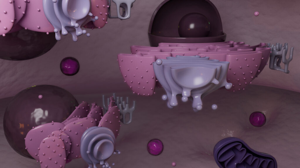
Osteoclast: 3D Render

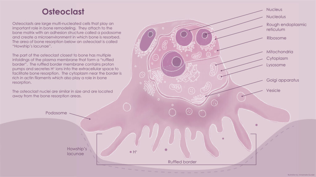
Osteoclast: Class PPT Slide

04: Major Histocompatibility Complex (MHC) Presentation

Project Overview:
The objective of this assignment was to create a didactic illustration for educational purposes. This illustration would be used in a textbook for an undergraduate Immunology class.
Intended Purpose:
This illustration is intended to teach undergraduate students the two different pathways for antigen presentation by major histocompatibility complex (MHC) class proteins. It depicts the endogenous pathway with MHC Class I presentation and the exogenous pathway with MHC Class II presentation. After viewing this illustration, students will be able to describe the pathways by which peptides are broken down within a cell for presentation by major histocompatibility complex (MHC) class proteins. Students will also be able to differentiate between the MHC class I pathway and the MHC class II pathway and explain the differences between the two.


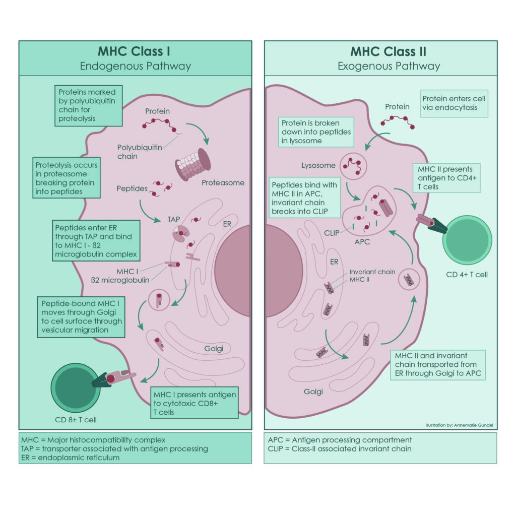
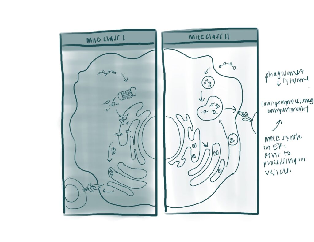
MHC Presentation: Final Image

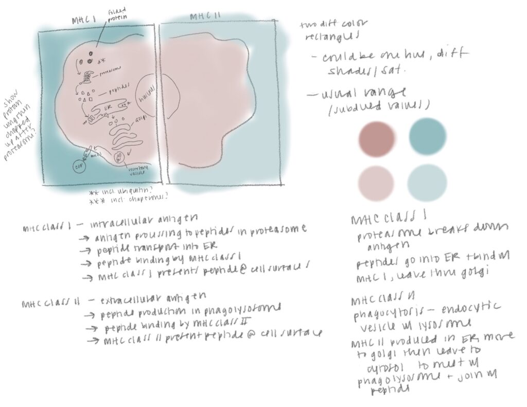
MHC Presentation: Detailed Sketch

MHC Presentation: Thumbnail

05: Medical Legal - Arthroscopic Bankart Repair
Project Overview:
The objective of this assignment was to create a medical legal exhibit based on a sample legal case.
Intended Purpose:
This exhibit is intended to support expert testimony and summarize the main steps in an arthroscopic repair of a Bankart lesion. The three major steps of the surgery are depicted, as well as references for the scapula location and position of the patient during surgery. After viewing this illustration the jury will be able to describe what a Bankart lesion is and how it is repaired with arthroscopic surgery.


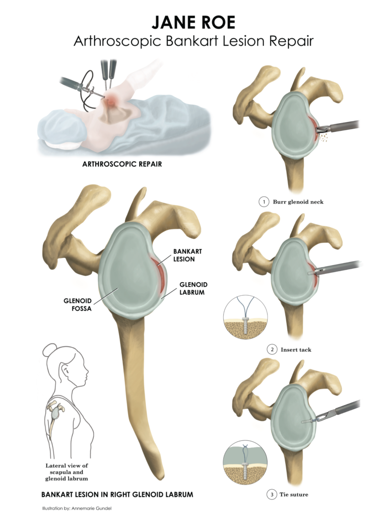
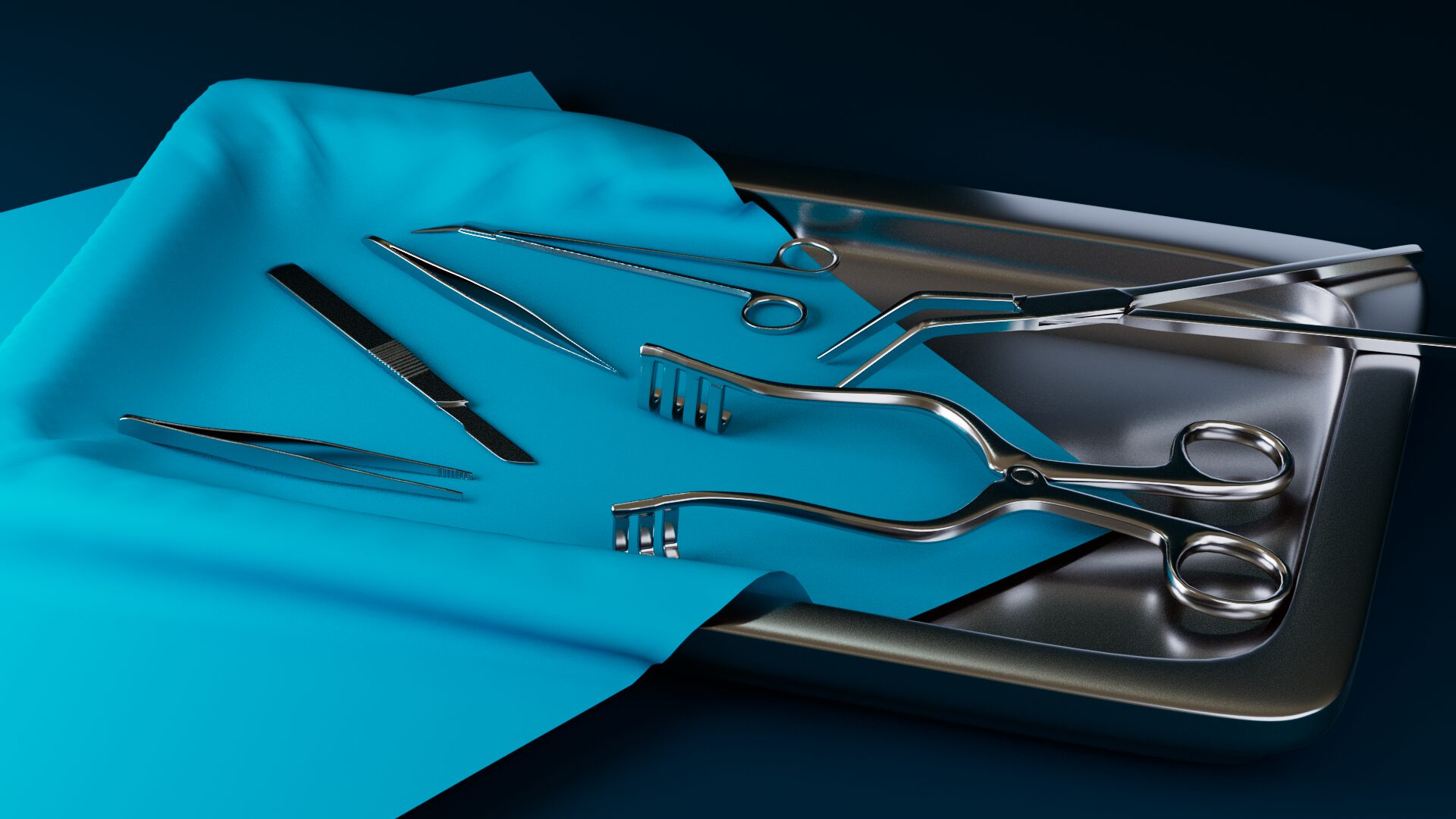
Bankart Repair: Final Exhibit

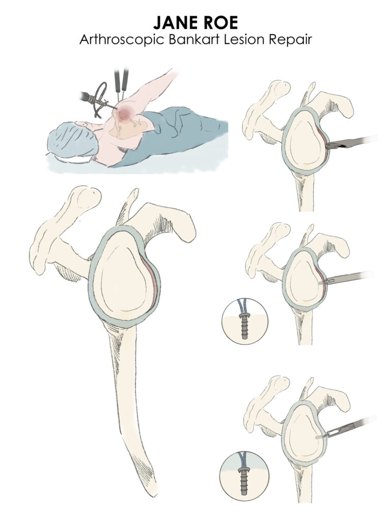
Bankart Repair: Initial Exhibit Sketch

06: Additional Work
- All
- 2D Illustration
- 3D Stills & Animation